 JPN
JPN


人生のすべての段階で、美しくなるのは私たちの権利であり、必要です。各患者様が自信と笑顔を取り戻すことを助けることは、すべての歯科医の願いです。今日のケースは、ポーランドのDr. Pawel Szuba-Paszkiewicz による事例で、86歳の女性に全面的な審美上顎復元を行ったことです。
ケースプロファイル
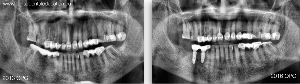
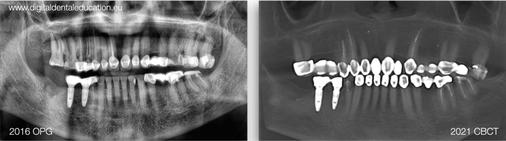
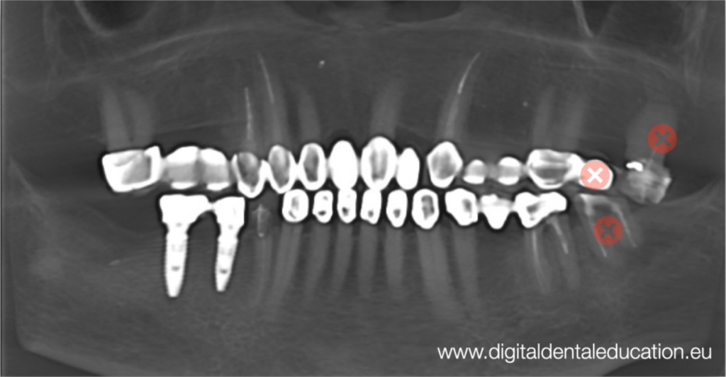
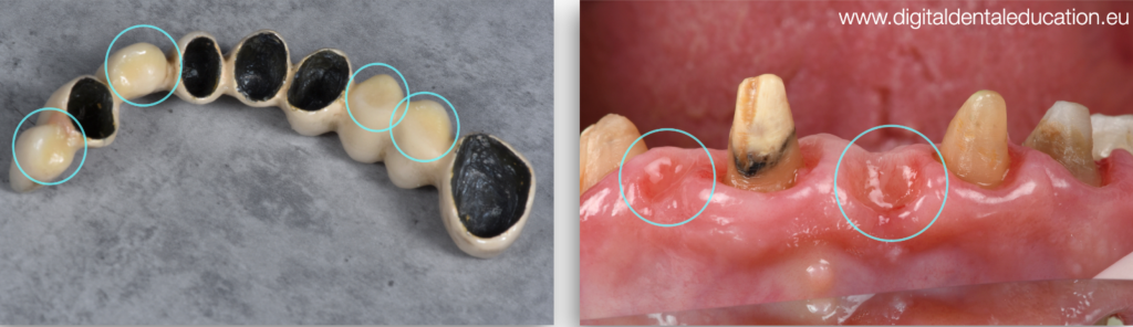
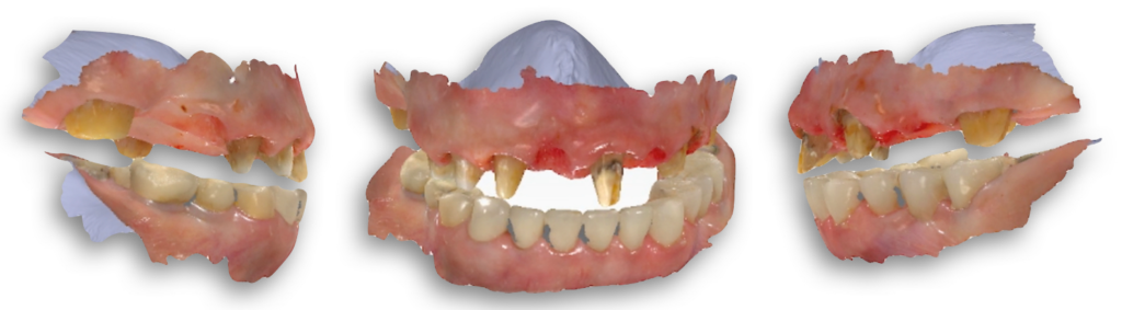
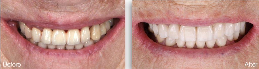
こちら患者は86歳の女性でした。患者は過去に何度も治療を受けていました。初期の年代では、下顎の後部の歯にインプラントが施され、別の病院で全上顎が再建されました(図1、図2)。しかし、時間のたちにつれて、密閉されていないブリッジや変色したブリッジが審美的また機能的な不快感を引き起こし、上顎のメタルーポーセレン破裂し、取り換える必要がありました(図3)。
診断
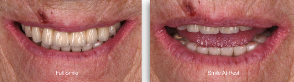
検討の結果、TMD障害、筋肉痛、咀嚼機能の不全、歯ぎしり、異常なスマイルライン、グループ機能、OVDの低下、摩耗はありませんでした(図4、図5)。患者の期待は、固定義歯による話し方と咀嚼性能の改善ができるとともに、上顎歯の色を明るくなれるし、より多くの歯を露出することでした。患者の年齢と下顎修復の良好な適応性および状態を考慮した上、歯医者が今の状態をそのまま保つことにしました。
治療前のDSD
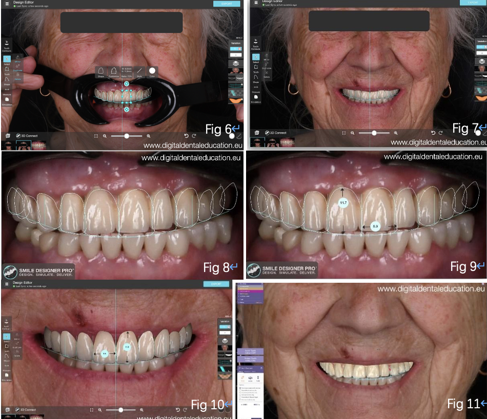
まず、歯科医は患者の高解像度の写真を撮り、歯科ソフトウェアでDSDを行いました。全面的なコミ ュニケーションを進んだあと、患者は歯の形の審美性に満足し、歯科医はさらなる治療を進めました(図6、図7、図8、図9、図10、図11)。
治療過程
歯科医は患者の口から古いブリッジを取り外しました。幸いなのは、ブリッジを丸ごとひとつの取り外し方法が見つけ緩和処理をしました、適切な歯肉生成と将来ポンティックの作成、適切な歯肉表面が形成されるまでの長期的な一時使用のものとしてそれを調整し、使用する方法がありました(図12、図13)。
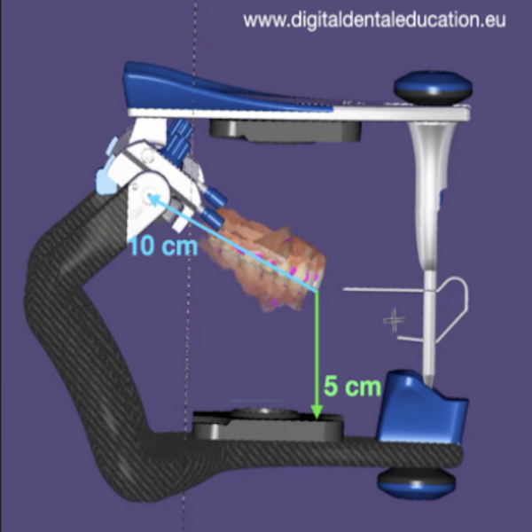
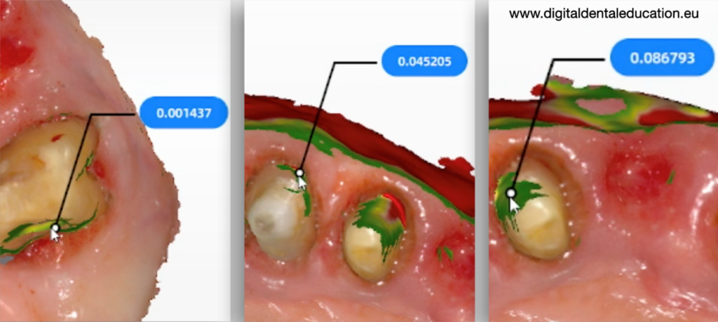
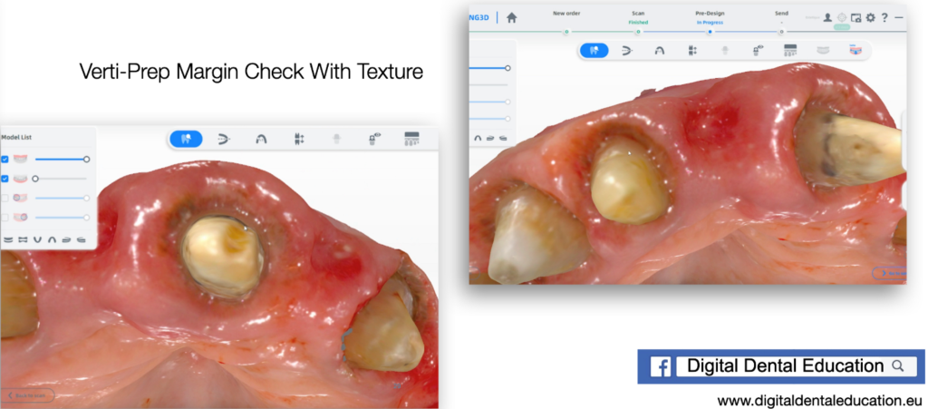
治癒の三ヶ月後(図14)、歯科医は歯の調整を精緻化し、最終修復のための口腔内スキャンを取ることにしました。彼はアンダーカットツールでアンダーカットをチェックし(図15、図16、図17、図18)、スマイルデザインと適切なバーチャル咬合のポジショニングのために写真とビデオによる手順を提供し、これはPhoto-DFA Workflowと呼ばれる歯科医師オリジナルの方法です。
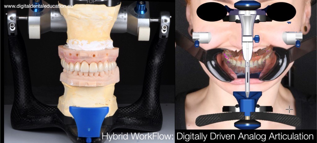
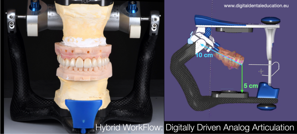
この方法は、審美と機能的な傾斜設定のためのフェースボウ概念と、回転軸の位置決めのためのDFAを組み合わせたものです。患者の確実に傾いた肖像写真を使用し、歯科医は3D口腔内スキャンデータを2D写真上で整列する手順を開発し、Kois DFAのアーテ ィキュレーションルールを使用し、仮想アーティキュレータ内のスキャンの適切な位置を達成することができました(図19、図20)。


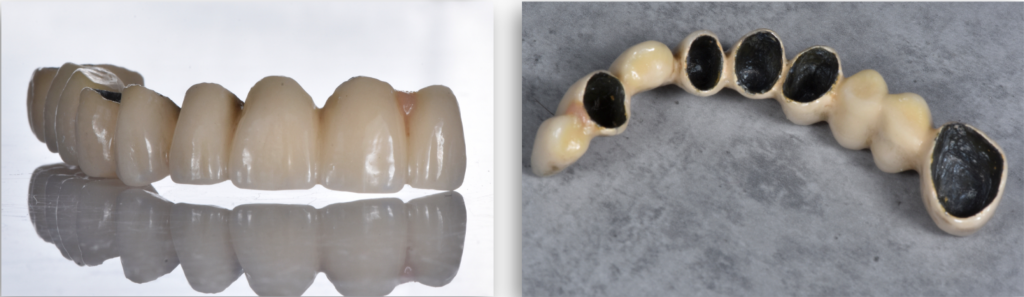
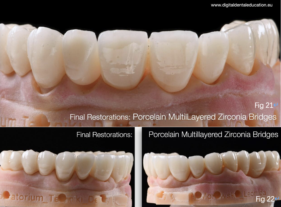
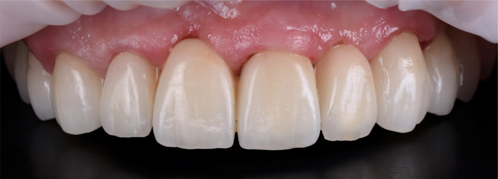
スキャンデータは3Dプリンターに送信されて印刷され、患者が確認したスマイルデザインに従って、歯科医は最終的な多層ジルコニアブリッジを作成しました。それはプリントしたモデルとよく合いました(図21、図22、図23、図24)。
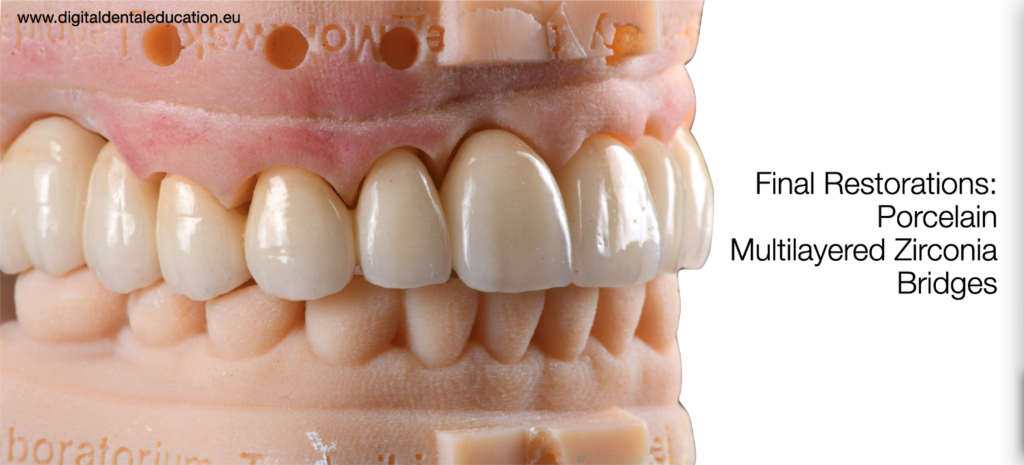
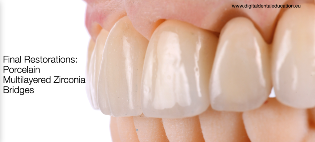
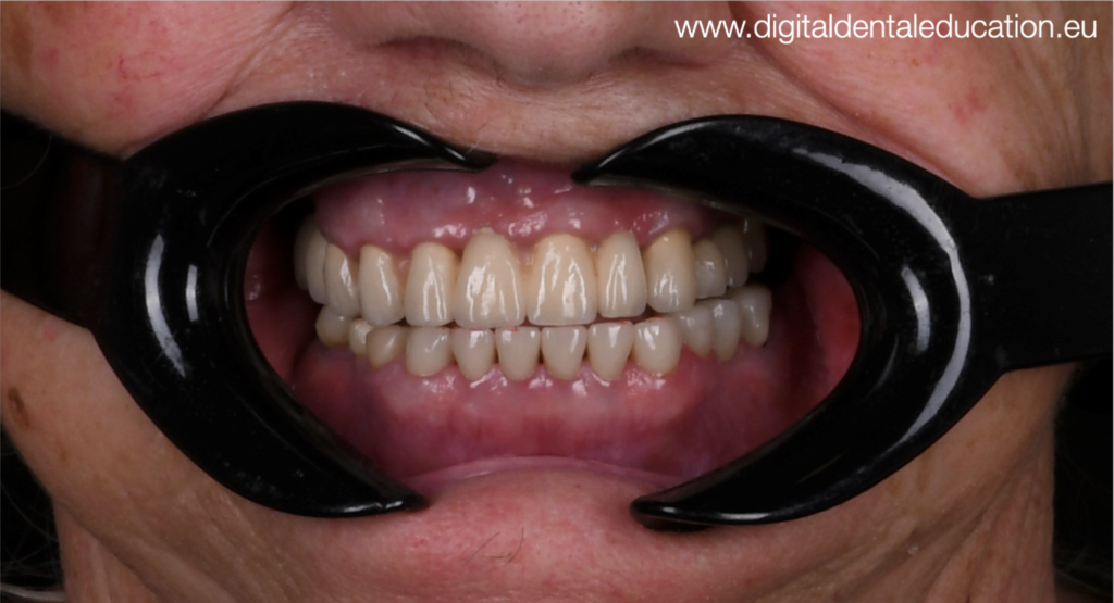
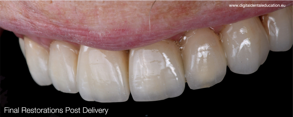
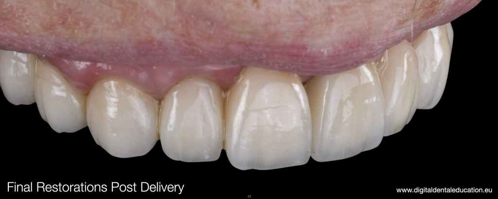
最終的な修復物は口腔内にセメントで固定されました。
歯科医のコメント
このケースにはAoralscan3という重要なスキャナーが使用されました。1分でスキャンを完了することができ、ジルコニアの破裂、亀裂、剥離などの合併症を避けるためには、大きなスパンのブリッジに対する高精度と正確性が不可欠です。
このアプローチは予約の回数を減らせます、通常には三回 で完了できます- 1.準備; 2.試適(常に必要ではありませんが、通常は患者は試適で新しい歯を受け入れると、すぐにそれらをセメントで固定できます);そして3.最終的な配達。もちろん、最終的な調整と詳細な検査とチェックアップのための追加の予約があるかもしれません。
歯科医Pawel Szuba-Paszkiewiczについて

Pawel Szuba-Paszkiewicz は 2004年、ヴロツワフ医科大学歯学部を優秀な成績で卒業しました。CAD /CAM補綴、インプラント補綴、フルマウスリハビリテーション、低侵襲審美歯科の分野で複雑な医療処置の治療を行っています。現在、彼の専門的な関心は、完全なデジタルワークフローを用いたデジタル補綴学とインプラント補綴学に集中しています。彼は、デジタル補綴処置中のティッシュ・リトラクションの革新的な手順であるEndo-Resto Single Visit Chairside Restoring Workflowや、革新的で高速かつ複雑なSmile Design & Delivering Protocol (SDP)、”Photo-DFA “ワークフローを開発し、導入しています。(図29)