 ENG
ENG


With the launch of the Aoralscan Elite, the world’s first intraoral scanner with photogrammetry, the accuracy of all-on-X implant cases has reached new heights. Increasingly, dentists are incorporating this scanner into their daily implant workflows and finding it extremely useful, as it eliminates the need for a verification jig and transforms their clinic procedures.
Today’s case comes from Dr. Hong Chang, an expert in implants with years of experience. Dr Hong is the founding partner of Experteeth Dental Group, which owns and operates 34 dental clinics as well as having its own Lab, Supplies and Academy in Australia. Dr Hong holds Diplomate status from the American Board of Oral Implantology and the International Congress of Oral Implantology.
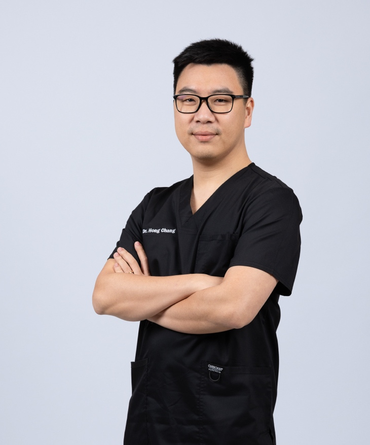
Fig 1: Dr Hong Chang: BDSc, MSc, DABOI, DICOI, FASID, FICD
Case Profile
A male patient who suffer from multiple missing teeth in the upper jaw for many years, affecting his aesthetics, function and phonetics. He visited Experteeth Dental seeking for help. He would like to rebuild the upper jaw to improve the masticatory function, facial appearance, and improve the life quality.
Initial Examination and Treatment Plan
Upon patient’s arrival, he expressed he want to get a stable, fixed restoration that would last for long term usage. So Dr. Hong Chang first took a CBCT scan to check the details, especially the bone conditions.
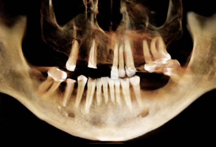
Fig: 2: The panoramic film revealed several a failing upper dentition, with periapical lesions associated with tooth #15, a retained root with tooth #13, and heavily restored teeth #21 and #22. The maxillary sinus in quadrant 1 showed pneumatisation.
Taking into account the treatment outcome, long-term stability of the restorations, the patient’s age, cost, and recovery time, decision was made to retain teeth 23, 25 and 26, while extracting all of the remaining upper teeth. A single implant is planned at 24 position, and a series of 4 implants are planned to restore the remaining teeth. Due to the pneumatisation of the sinus present in quadrant 1, an all-on-X style solution with a tilted implant at the 16 position is planned. This approach minimizes surgical trauma while maintaining long-lasting stability. The focus of this treatment will be on the upper jaw, with the lower jaw addressed in the future.
Treatment Process
Extraction was performed first. After a time period of wound recovery, the patient came back, a series of record was taken to enable formulation of a digital wax up.
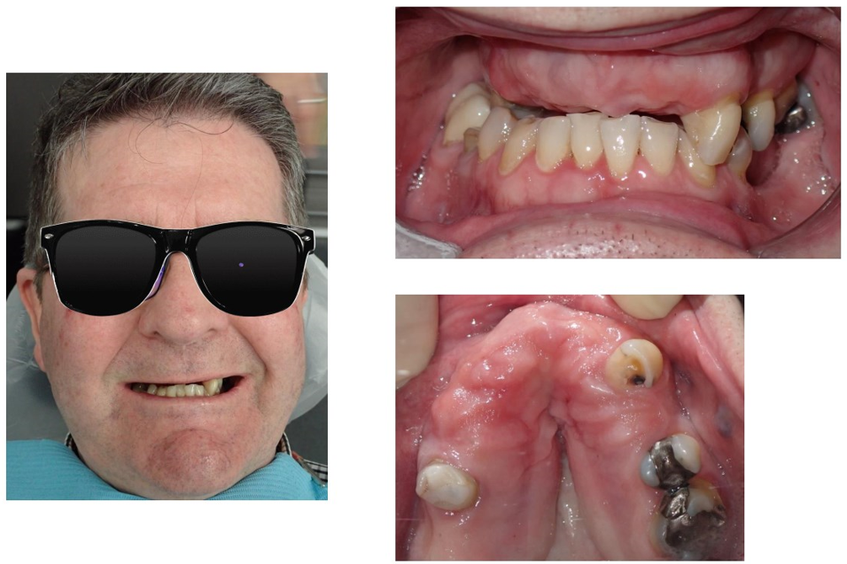
Fig 3: The face photo from front sight view.
Fig 4: The lower jaw from front sight view.
Fig 5: The upper jaw from occlusion sight view.
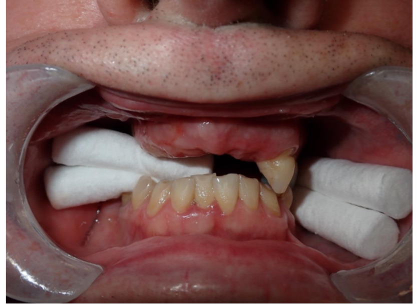
Fig 6: Record the vertical demension by Shining 3D Elite Scanner with the aid of cotton roll.
Immediate loading is planned for this case. During the implant surgery, Dr. Hong Chang placed five implants and immediately captured intraoral data using the Elite’s coded photogrammetry scanbody workflow for four of the implants.
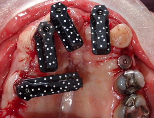

Fig 7, 8: Installed the coded photogrammetry scanbody and scanned the data. Even if there is blood intraorally, Elite’s advanced algorithm can capture the data without any problems.
After capturing the coded photogrammetry scanbody data, the software back calculates and virtually places the chosen scan body library onto the calculated implant positions. This process can be done in the intraoral scan software or on the cloud. Many implant brands have been integrated into Elite’s coded scanbody workflow. For implant brands not listed in our software, users can locally import the library into the software themselves if they confirm that the coded scanbody fits properly with their MUA.
The lab performed a smile design and fabricated the temporary restorations for the patient.

Fig 9, 10: The Technician designed the restorations in exocad based on the intraoral scan data as well as the original photos.
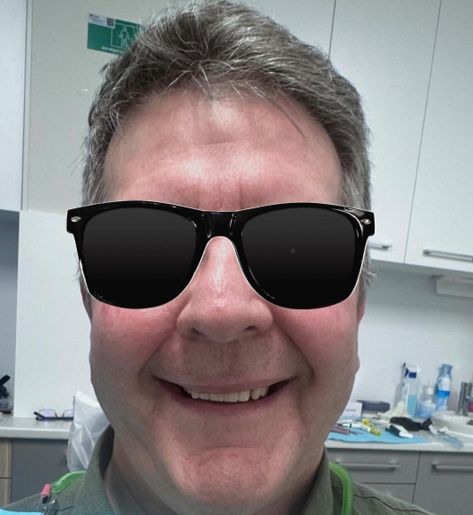

Fig 11, 12: The temporary restorations were delivered to patients on the same day, achieving a passive fit, and the trial process went very smoothly.
After several months, after osseointegration is confirmed, we started permanent restoration construction. The patient returned, and Dr. Hong Chang scanned the data again. The lab technicians made slight adjustments to the original design to make it more refined and suitable.
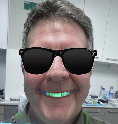
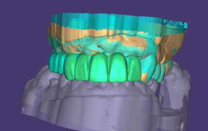
Fig 13, 14: The second design in exocad.
Final Restoration and After Treatment
The final restoration was made of Zirconia on a Toronto bridge, which fits well in the patient’s mouth without any rocking or adjustments. The final teeth wearing process was very smooth and only took minutes.
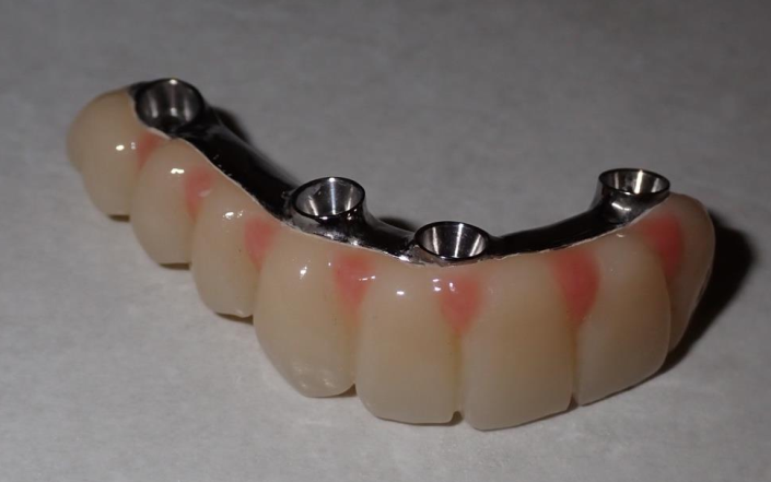
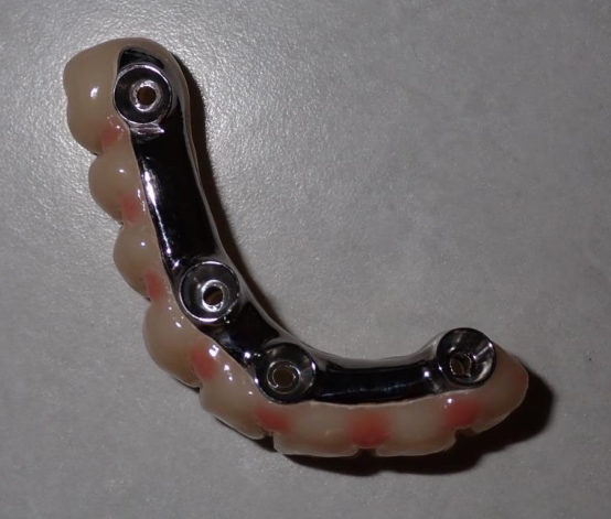
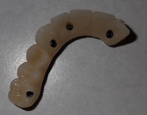
Fig 15,16,17: The final restorations
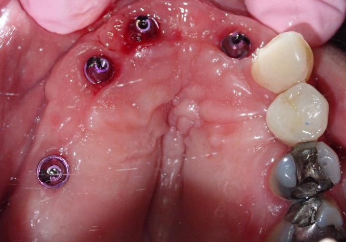
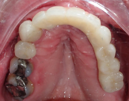
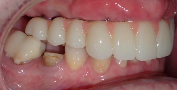
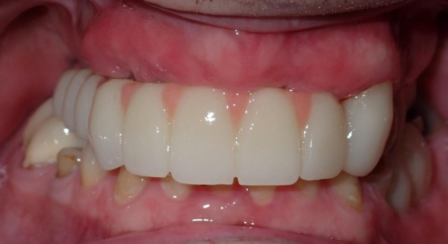
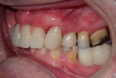
Fig 18,19,20,21,22: Intraoral view during and after treatment
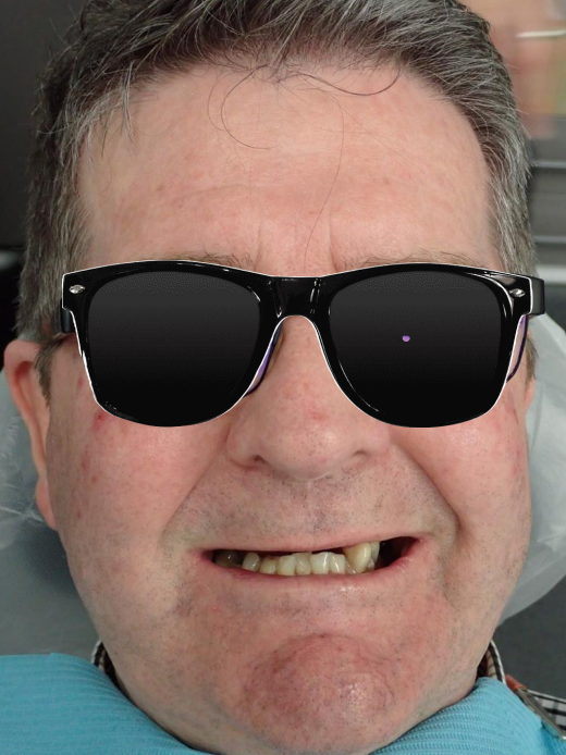
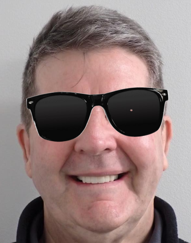
Fig 23, 24: The comparison before and after the treatment

Fig 25: Final OPG showing excellent fit
Comments from Dr. Hong Chang:
As one of the earliest users of Elite in Australia, we are super excited to have the chance to try out this device, which combines intraoral scanning and photogrammetry into a two-in-one system. We’ve tested it on some of our initial cases, and it works really well, very fast. With its accurate scan data, it delivers precise restorations that fit perfectly, previosuly only possible with a separate photogrammetry scanner. The two-in-one solution is truly a revolutionary device and we are super excited to use it on more patients in the future.