The advent of digital technology has revolutionized the field of dentistry, transforming the way in which dental professionals approach their workflow and practice. With the rise of cutting-edge intraoral scanners, face scanners, and printers, these innovative tools have become essential players in modern-day dental treatment.
Today’s case comes from the experienced Dr. Golovanova Polina Andreevna, a highly skilled dentist. She has devoted her career to providing exceptional dental care to patients in need. Dr. Golovanova is passionate about helping her patients achieve optimal oral health and restoring their beautiful smiles. Thanks to the innovative technology provided by SHINING 3D, Dr. Golovanova can now offer even more efficient and effective treatments to her patients.

Fig 1: Dr. Golovanova Polina Andreevna
Case Profile
A middle-aged woman, aged 56, has been experiencing the distressing effects of missing posterior teeth for quite some time now. This unfortunate condition has made it difficult for her to chew properly and enjoy her meals. Seeking relief from this persistent problem, she decided to seek professional help and approached Dr. Golovanova with a request for treatment.
Treatment Process for Implant Restoration
After conducting a thorough examination and engaging in comprehensive communication with the patient, it was determined that two implants were necessary to be placed in the #36 and #37 areas of the Dentium Superline system. The surgery was successfully completed, and after two months, we were pleased that the emergency profile was good, and the wound has fully recovered. We can move forward to make the upper restorations.
First, Dr. Golovanova carefully examined the patient’s dental implant to ensure that it was properly integrated with the surrounding bone tissue. To do this, she took a panoramic radiograph which provided a detailed view of the implant and its surrounding structures.
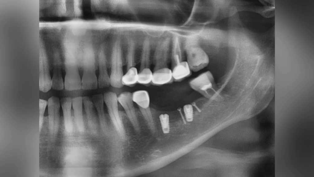
Fig 2: A detailed view of the implant and its surrounding structures
Next, an intraoral scan was performed using the state-of-the-art product Aoralscan 3. This advanced intraoral scanner allowed for incredibly accurate data capture within just one minute!
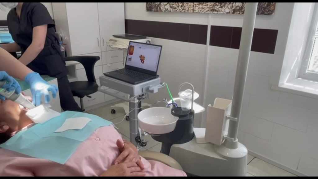
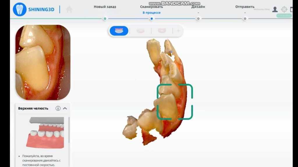
Fig 3, 4 Using Aoralscan 3 to capture intraoral data
Once all necessary data had been collected and analyzed, the data had been imported into the exocad to design.
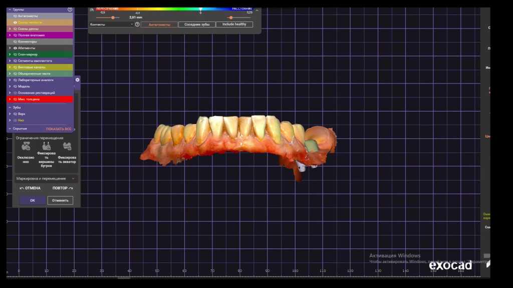
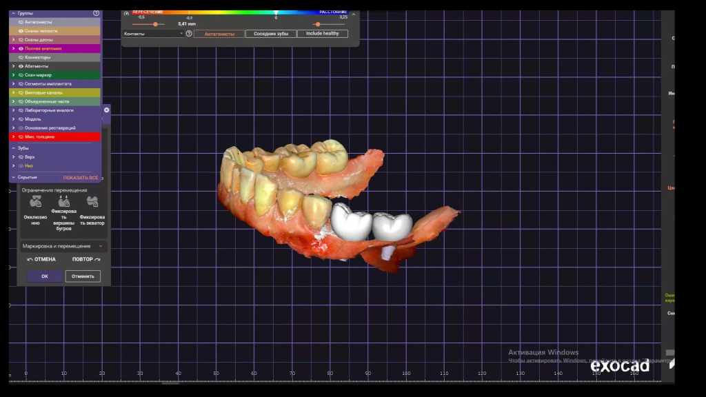
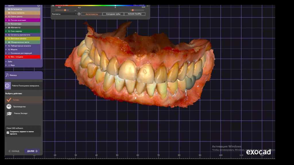
Fig 5,6,7: Design data in exocad
After the design process, the dentist sent the files to a lab for further fabrication. At the same time, the dentist printed a model internally with AccuFab-L4D using resin DM12. To ensure that the models were solidified properly, they used Fabcure curing machine which helped in achieving accurate model. Once completed, two restorations were tested on the printed model to check if there were any unsuitable areas or issues such as occlusion problems or improper morphology. If any issues arose, adjustments would be made immediately.
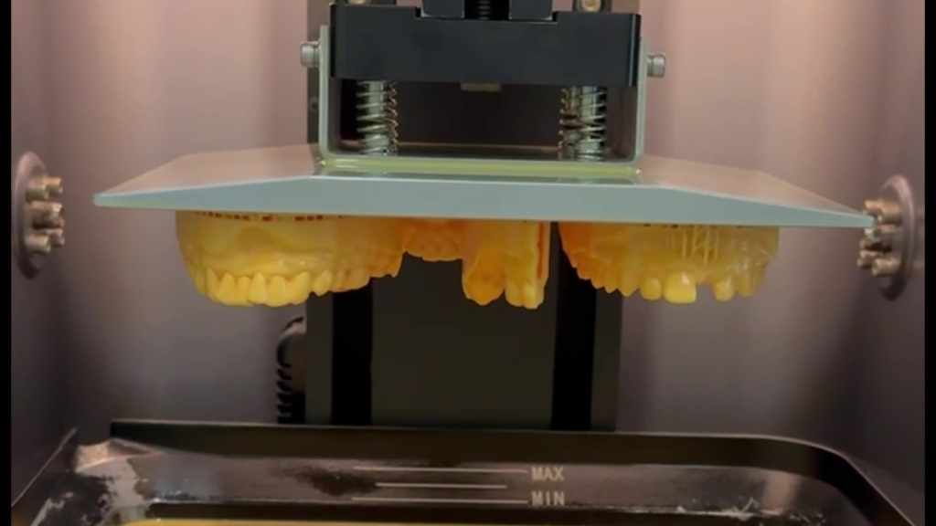
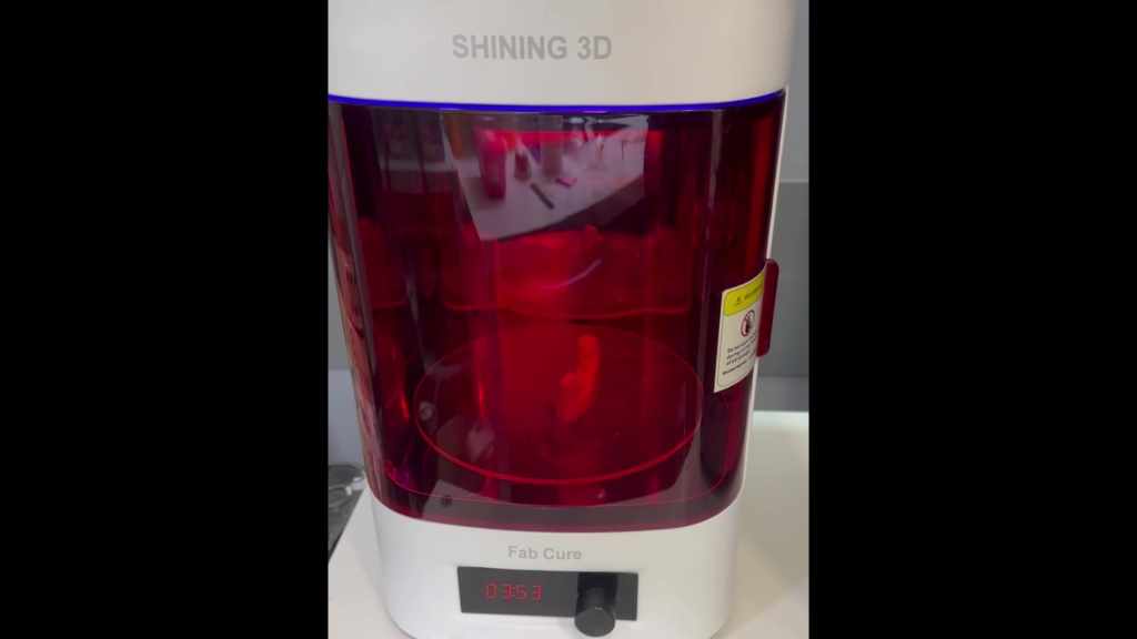

Fig 8,9,10: Printed a model with AccuFab-L4D using resin DM12
After the zirconia restorations were ready, the dentist proceeded to fix them into the patient’s mouth. After a standard cement process (Fig 11), the restorations were securely positioned in place, ensuring maximum comfort and functionality for the patient. As Fig 12 and Fig 13 showed, the zirconia restorations were successfully fixed into the patient’s mouth.

Fig 11: A standard cement process

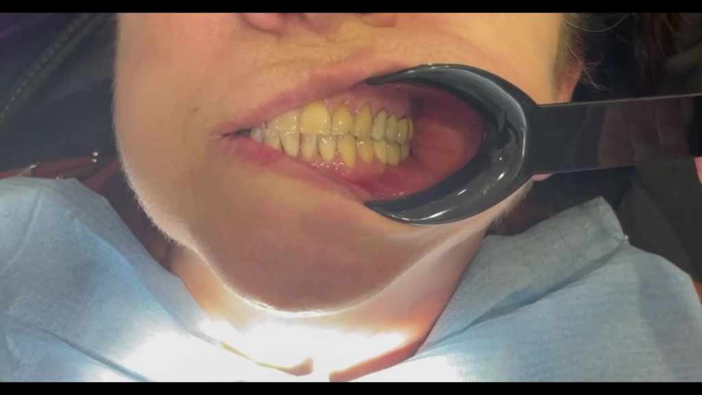
Fig 12,13: The zirconia restorations were successfully fixed into the patient’s mouth.
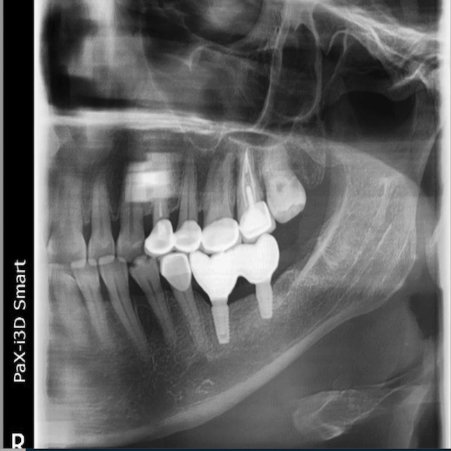
Finally, the dentist took a panoramic radiograph to assess whether the restorations were placed in the correct position.
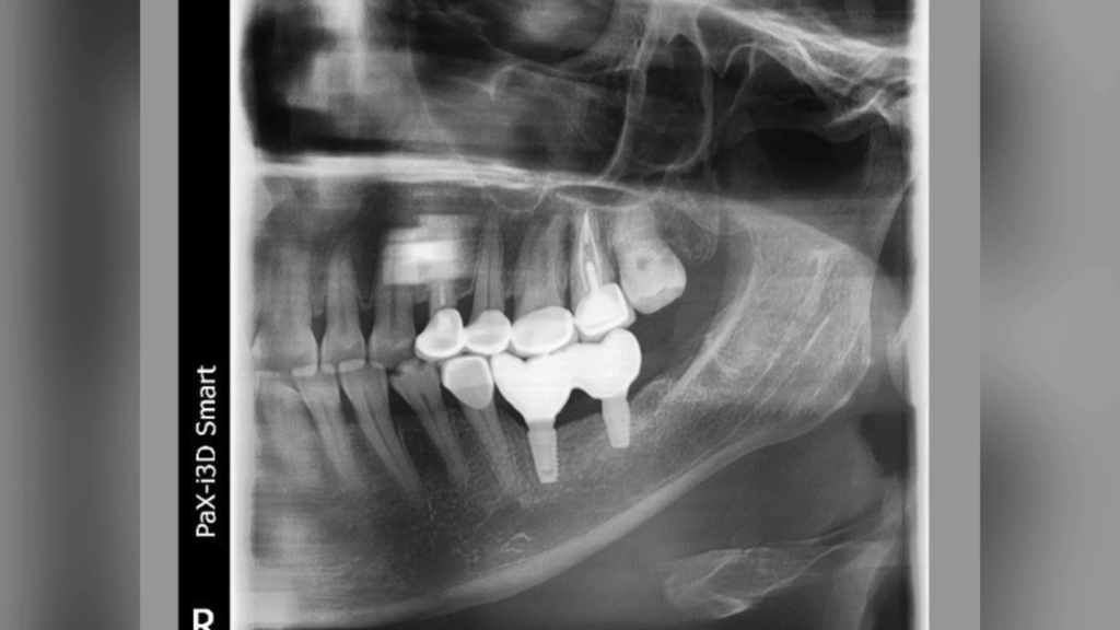
Fig 14: The restorations were placed in the correct position.
Comments from Dr. Golovanova Polina Andreevna
I’ve been using the Aoralscan 3 intraoral scanner and AccuFab-L4D for years, these products had helped a lot to give a more efficient and effective treatment to my patients, and I am happy to go digital with SHINING 3D.
 ENG
ENG


