 ENG
ENG


Since the launch of the Aoralscan Elite, dentists have been exploring its potential in all-on-X implant cases using photogrammetry technology. However, the Elite isn’t just great for all-on-X edentulous implants, it also performs exceptionally well in normal implant cases.
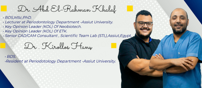
Today’s case comes from Dr. Abdelrahman Khalaf and Dr. Kirollos Hany (Fig1). Their study highlights how the Aoralscan Elite, together with MetiSmile, can significantly enhance the quality of dental restorations in the anterior area.
Abstract
This article explores the combined use of digital implant surgical guides and Guided Bone Regeneration (GBR) in the aesthetic restoration of missing anterior teeth with insufficient bone volume. The focus is on the precise treatment process, from 3D scanning and design, to bone augmentation, and finally, the restoration with advanced Aoralscan Elite and MetiSmile.
Before Treatment
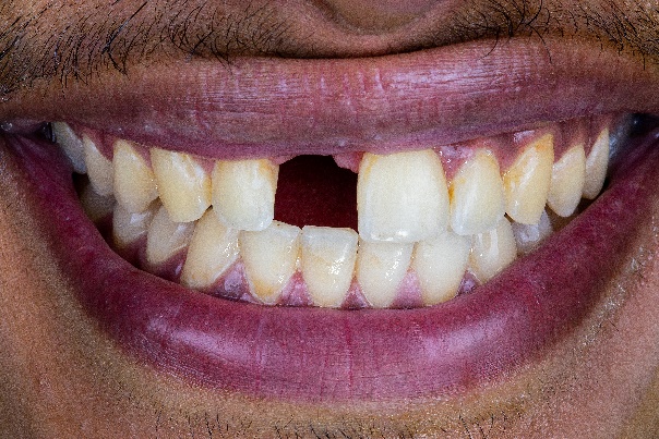
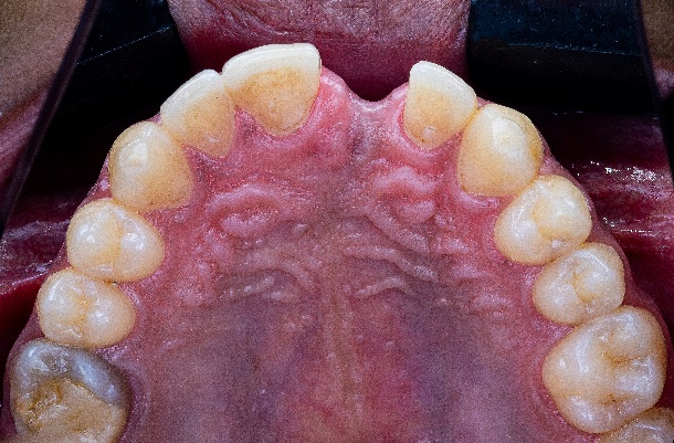
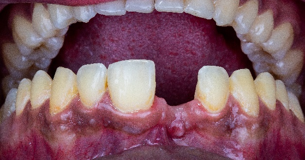
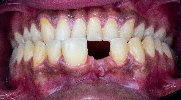
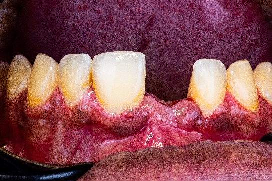
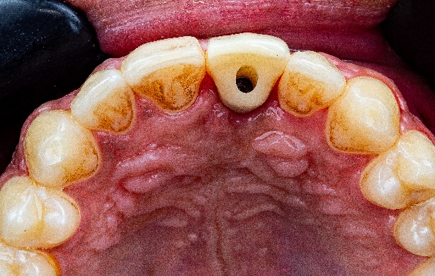
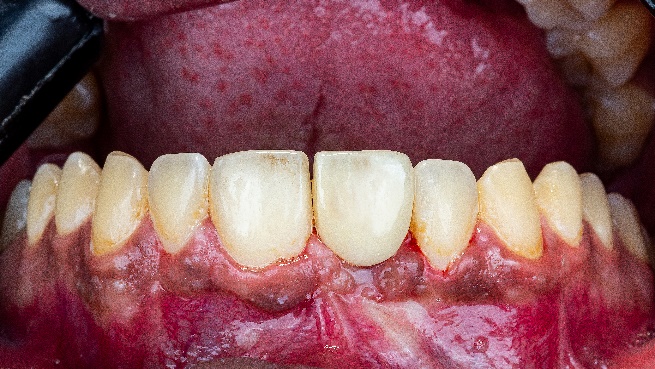
The patient, a 35-year-old male, sought treatment six months after losing his anterior tooth (21) due to trauma. Clinical examination revealed soft tissue depression in the edentulous area, while CBCT imaging showed insufficient horizontal bone width (<4mm) in the region, with normal vertical bone height, adjacent teeth, and occlusal relationships, the patient has high aesthetic demands. (Fig2,3,4,5)
Treatment Process: Aesthetic Analysis and 3D Imaging Acquisition by Aoralscan Elite and MetiSmile
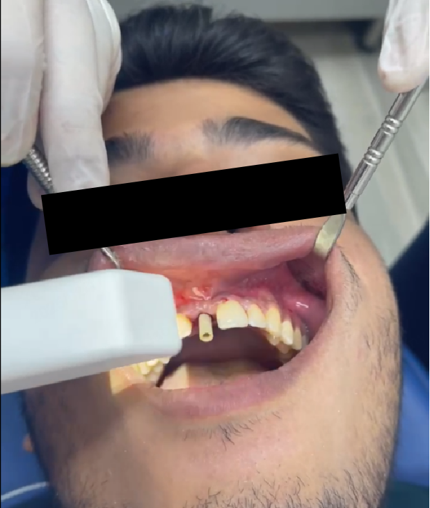
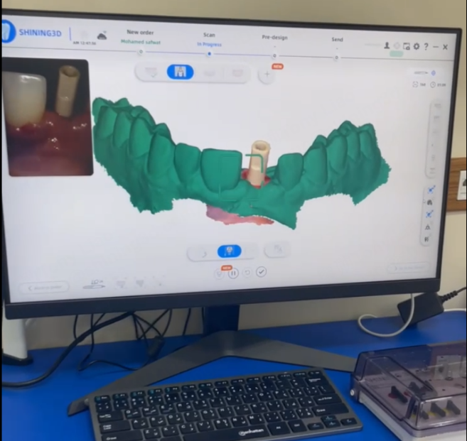
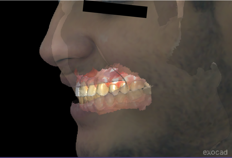
The latest intraoral scanner, the Elite from Shining 3D, was used to quickly capture stl data of both the upper and lower jaws, as well as gingival morphology and proportions relative to adjacent teeth (Fig 6). Additionally, the MetiSmile face scanner from Shining 3D, combined with digital smile design, was utilized to determine the morphology and incisal edge position for the future restoration (Fig 7).
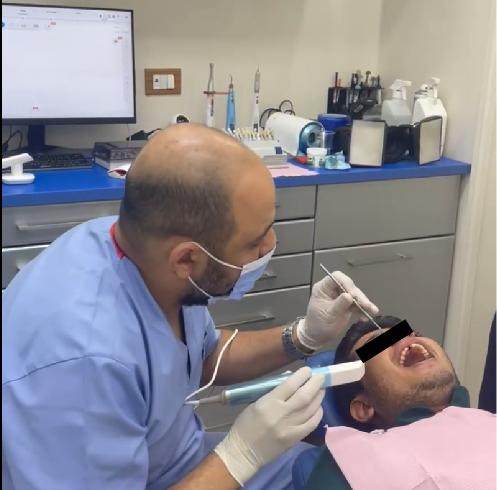
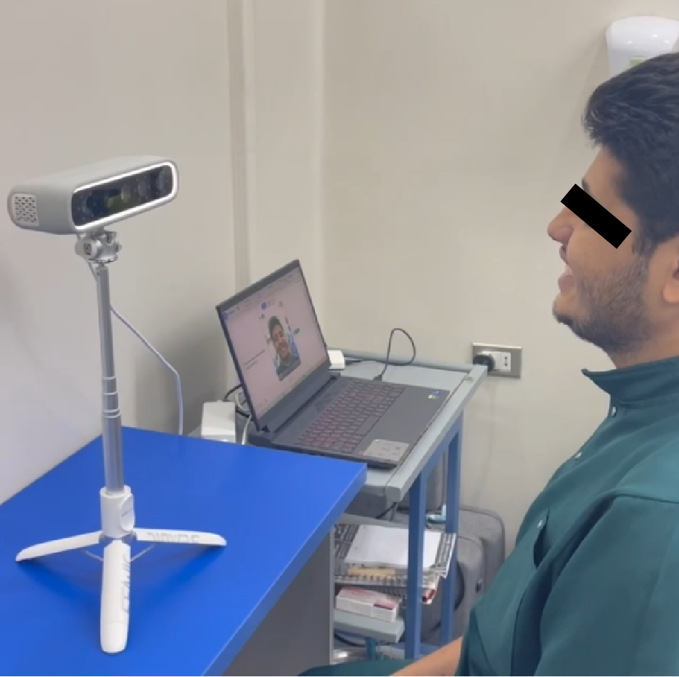
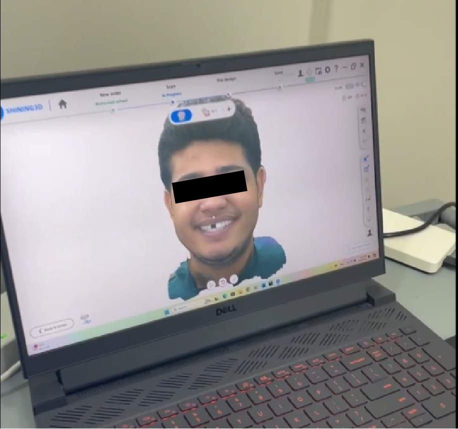
Fig 8: The scanned face shows in the software.
Treatment Process: Virtual Implant Planning
CBCT and intraoral scan data were imported into implant planning software to simulate the 3D positioning of the implant, ensuring a depth of 3mm from the future restoration’s gingival margin and a ≥2mm bone plate on the labial side. The software then generated a tooth-supported implant guide.
Treatment Process: Guided implant Placement and Minimally Invasive Bone Augmentation Surgery
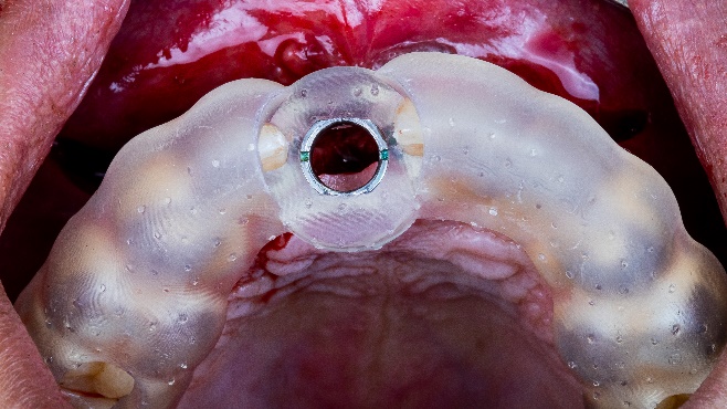
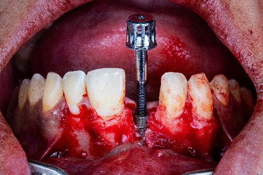
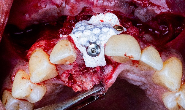
The surgical guide, made from transparent resin, was printed using Shining 3D’s AccuFab-D1s printer. After positioning the guide, the bone grafting area was precisely identified with its assistance. Implant socket drilling was carried out step-by-step under the guide’s direction, and the implant was successfully placed with excellent primary stability. (Fig 9, Fig 10)
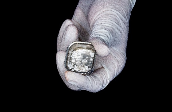
The bone material was implanted and covered with a non-resorbable titanium-reinforced Polytetrafluoroethylene (PTFE) membrane, which served as a barrier to prevent cell occlusion. The edges of the membrane were secured with a titanium nail(Fig 12,Fig 13)
Treatment Process: Second Phase Restoration
Six months after surgery, the evaluation of bone augmentation showed that the labial bone thickness had been restored to 5.5mm, meeting the conditions required for aesthetic restoration, and the osseointegration is good.
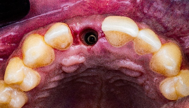
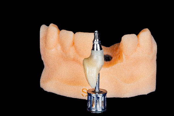
The scanbody is placed, and the Elite intraoral scanner is used to capture data for the design of the subsequent temporary crown. Since this is a single implant restoration, there’s no need to use IPG technology for determining the implant position (Fig 16, Fig 17).
Both the facial and intraoral data are imported into exocad for digital smile design. With the reference of the facial scan data, the technician can better understand the patient’s facial features and design the restoration’s morphology and smile line accordingly. (Fig 18,19,20,21)


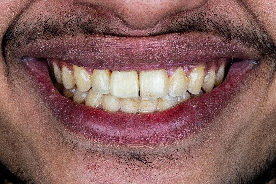
The temporary crown try-in showed a good margin fit, evenly distributed occlusal contact points, and the morphology was confirmed by the patient. Later, the final zirconia crown was delivered to the patient, who expressed great satisfaction with the final result. (Fig 22,23,24,25)
Dr. abdelrahman khalaf and Dr. Mohamed Khalaf’s comments on Aoralscan Elite
I bought the Aoralscan Elite for its good performance in all-on-X implant cases with IPG technology. What surprise me, this scanner is also good at the normal implant workflow as well as the other restoration cases. So I am confident to say, this scanner is an all-in-in scanner. One scanner for all the applications, I like to explore more this scanner with lots of upcoming cases.