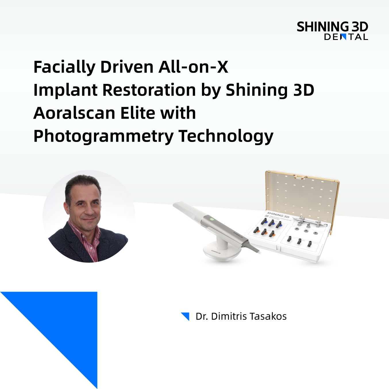
The accuracy of all-on-X implant impressions and the passive fit of restorations remain significant challenges in implant cases. The newly launched SHINING 3D Aoralscan Elite addresses these challenges effectively by introducing photogrammetry to implant dentistry. This innovative two-in-one device captures precise implant positioning through intraoral photogrammetry, while also obtaining soft tissue details via edentulous scanning using structured light.
Since its launch in September 2024, thousands of successful cases have been completed with this technology. Today, we highlight a case from Dr. Dimitris Tasakos, who combines cutting-edge digital techniques, such as intraoral photogrammery, facially-driven implant planning, and facially-guided aesthetic restoration technology, to achieve highly accurate full-arch restorations.
Dr. Dimitris Tasakos is a DSD Master and ICOI Fellow, renowned for his active involvement in lectures and hands-on seminars on Digital Dentistry. His expertise spans Digital Impressions, Digitally Guided Implantology, and prosthetic restoration of implants, with a particular focus on direct loading protocols. Dr. Tasakos operates a dental clinic in Athens, where he offers comprehensive treatment using advanced digital technologies.
Case Analysis and Treatment Planning
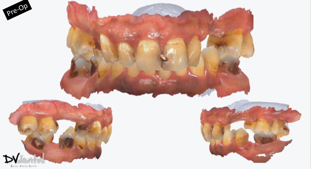
A 47-year-old male patient presented with severe resorption of the maxillary alveolar bone, as confirmed by X-ray examination. Significant caries were present on teeth #14, #11, #21, and #27, while natural teeth #15, #23, #24, and #25 have already been lost.
After discussing treatment options with the patient, it was decided to focus on the upper jaw first, retain only teeth #16, #17, and #26 in the maxilla. The remaining teeth will be extracted. Four implants will be placed at positions #15, #12, #22, and #25 to support a subsequent implant-supported bridge restoration.
After completing the treatment for the upper jaw, the patient will proceed with rehabilitation for the lower jaw at a later stage.

Data Capture using Aoralscan Elite and MetiSmile
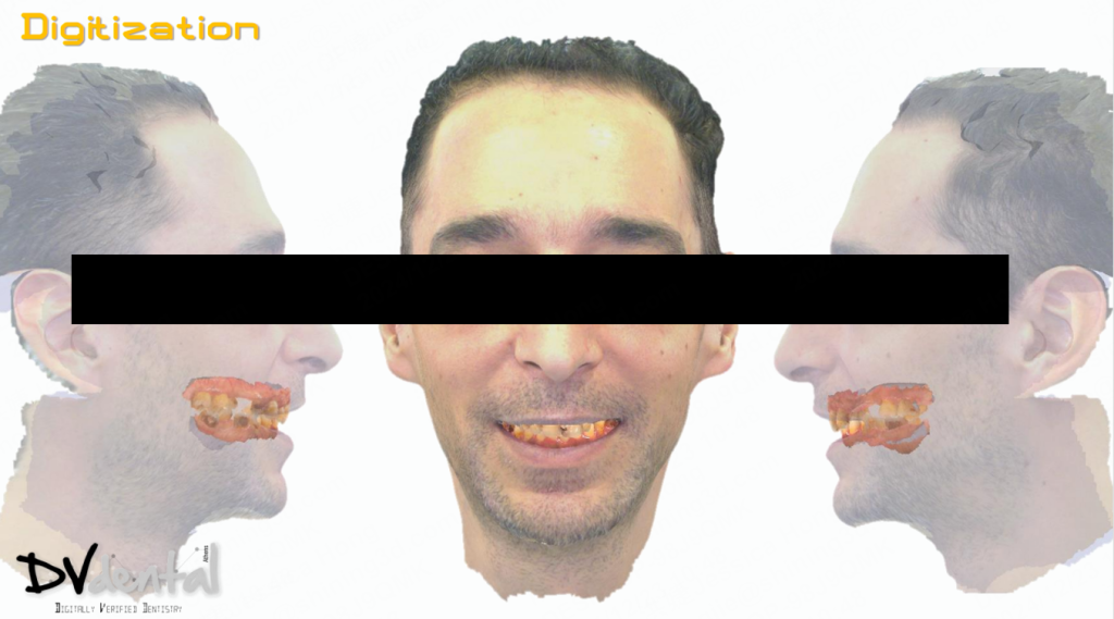
Using the SHINING 3D Aoralscan Elite to capture the upper and lower jaw accurately, while the Metismile face scanner gathers detailed facial data. After that, obtain CBCT data, and it can be seamlessly imported and aligned with both the jaw and facial data for precise implant planning and the design of a temporary bridge in the later stage.

Implant Surgery and Immediate Loading
The tooth-supported surgical guide was designed and manufactured, followed by implant surgery performed under its guidance. Post-suturing, the SHINING 3D Aoralscan Elite was used immediately for scanning. Thanks to its advanced photogrammetry technology, we were able to achieve highly accurate restorations with a passive fit for the temporary bridge during the first stage of immediate loading.


Second-stage Restoration
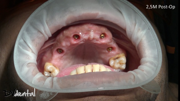
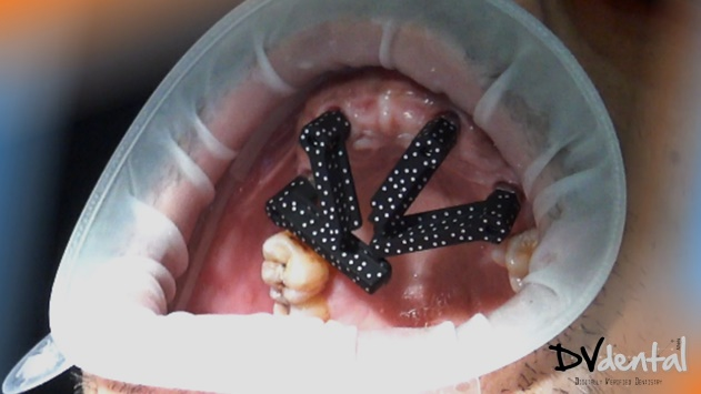
After 5 months of healing, the bone and implants were well-integrated, and the dentist was ready to proceed with the permanent restoration. The SHINING 3D Aoralscan Elite was used to capture data of the upper jaw soft tissue, lower jaw, and implant positions. To ensure proper fit of the coded scanbody, an X-ray was taken for confirmation. Upon completion of the scan, the coded scanbody data was converted into the corresponding implant library for final restoration planning.


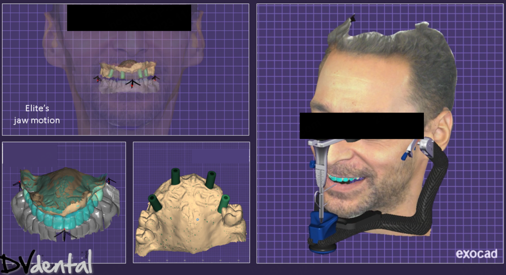
Using the face data to mount the model on the virtual articulator, design the permanent bridge based on the temporary restoration. Since dynamic occlusion data was previously collected with Elite, we can eliminate any interference during the occlusion adjustment process in exocad.
Upon trial fitting in the patient’s mouth, the permanent bridge seated passively on the MUA. X-ray examination confirmed that all margins are well-adapted. The shape of the teeth, midline alignment, and incisal edge position closely mirror those of the temporary restoration. The patient is satisfied with the esthetic outcome and is pleased with his new smile.


Dr. Dimitris Tasakos’s comments on Aoralscan Elite with IPG Technology
Having been in dental implant practice for many years, I can confidently say that the Aoralscan Elite has truly transformed my All-on-X implant workflow. With this device, I no longer have to deal with the cumbersome and complex traditional implant impression process. Instead, I can easily and accurately capture both the implant positions and soft tissue through scanning. I look forward to incorporating it into more cases in the future.
 ENG
ENG


