The implant reconstruction of anterior teeth in the upper jaw is not only important in recovering the masticatory system but also in retaining a natural aesthetic. Digital technology helps to simplify the process and reduce treatment time, especially the implantation in anterior teeth.
Today’s case comes from Dr. Wang Han and technician Zhou Xi, they come from Hangzhou Dental Hospital. Wang Han is a deputy director of physicians, the director of the Digital Medical Project, and a specialist in digital implantology. Zhou Xi is the manager of a digital technology centre and a dental medical technician.
Case information
- Patient’s information: 39-year-old female
- Chief complaint: the colour of the gingiva of the anterior teeth has been changing for years
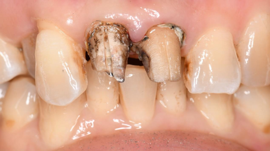
- Present illness: The patient was treated 10 years earlier with porcelain teeth. However, the gingival recession has progressively exposed the metal margin of the previous restorations. The patient came in for a visit and required treatment as this is a highly aesthetic area.
- Medical history: No history of allergies or any other disease.
- After clinical examination, it was determined that #8 and #9 should be further restored.
Treatment plan
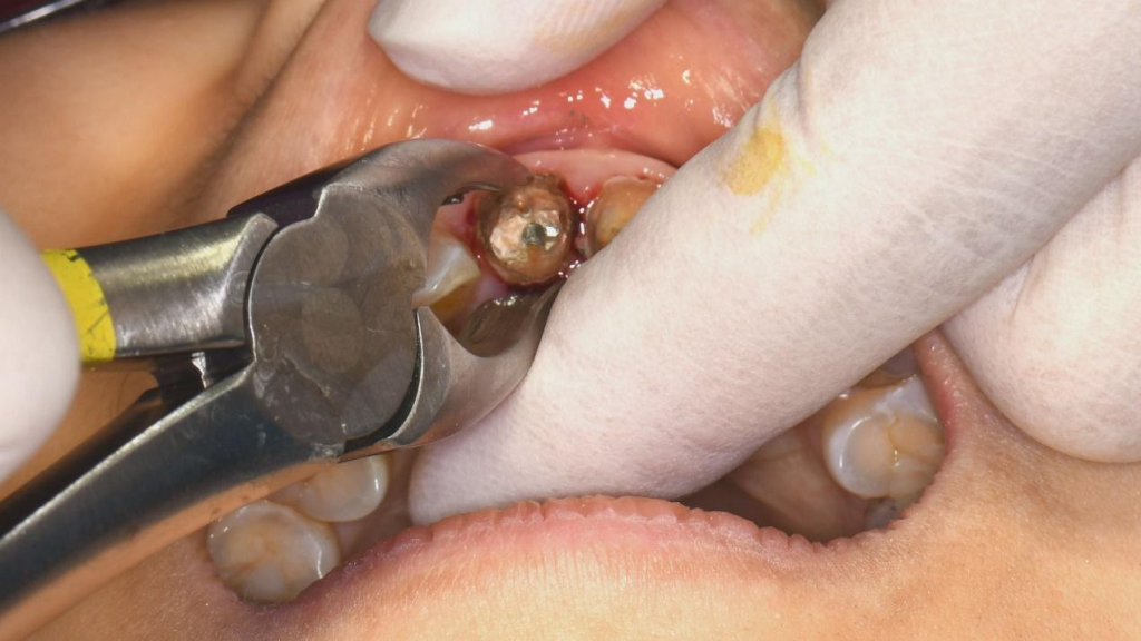
Remove previous crowns on #8 and #9, replace #9 with a new full porcelain crown, extract #8, and follow up the extraction with immediate implant surgery.
Before treatment
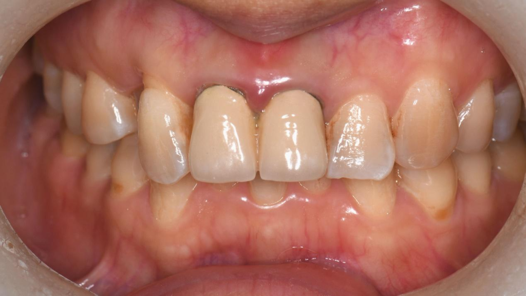
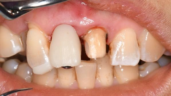
Examination: #8 and #9 are porcelain fused to metal crowns. There are suspensions at the edge of the prosthesis, and the margin of the gingiva was receding with dark colour, thin biotype, and gingival papillae were red and swollen (Fig. 1).
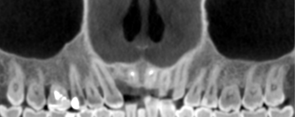
CBCT: After RCT and post and core treatment, the old post deviated to the middle third of the labial side of the tooth root. The width of the alveolar bone was 7.7 mm, and the labial bone plate was 1 mm. There was no obvious absorption shadow at the root tip of #9 (Fig. 2)
Data capture before operation
Intraoral scan data
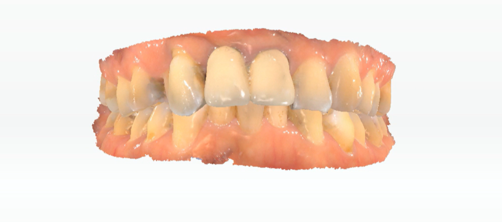
After determining the treatment plan, the dentist captured the patient’s intraoral data using the Aoralscan 3, quickly and accurately. Together with HD CBCT data, it was prepared for the design of a digital implant surgical guide (Fig. 3).
CBCT data
The design project
The implant surgery planning
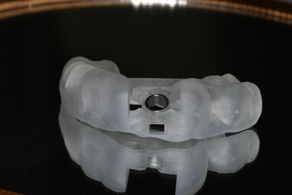
Using the combined CBCT and intraoral scan data, the three-dimensional site of the implant and surgical guide were designed to ensure the safety of the implant surgery and good positioning for an aesthetic restoration. Using this method, it is possible to account for the size of the extraction socket, the position and initial stability of the implant, as well as the very sensitive aesthetics of the anterior teeth.
Design the temporary teeth with exocad
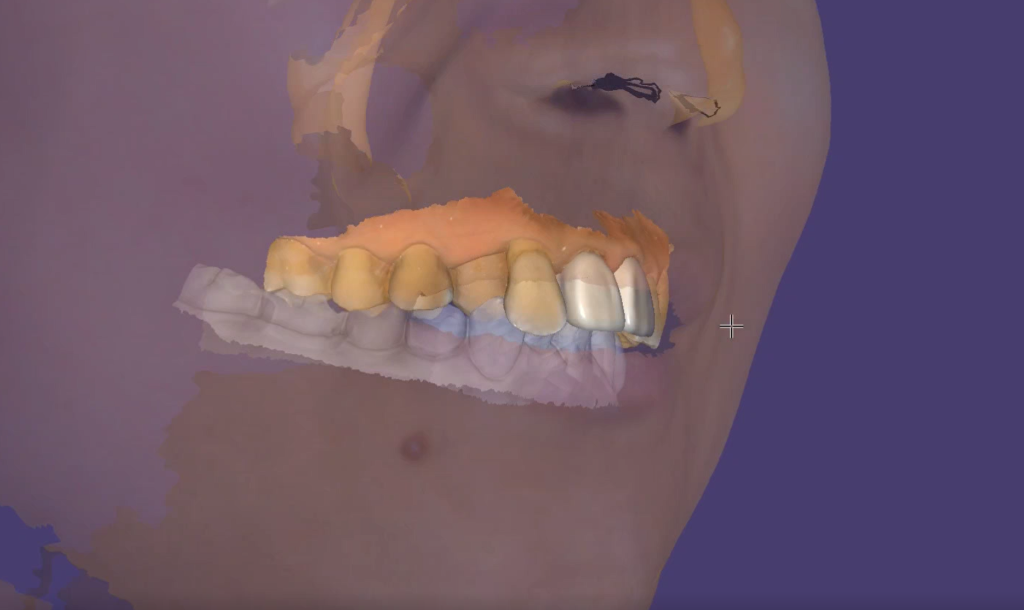
With face scan data and DSD, the temporary teeth were designed in the CAD software and created in advance to meet the patient’s needs and to streamline the process of restoring the teeth during the operation (Fig. 4).
Print the implant surgery guide plate
With the AccuFab-D1s from SHINING 3D, the digital surgical guide can be generated through automatic layout, support-adding and slicing within the dental software Accuware (Fig. 5). Temporary teeth were made before the surgery (Fig. 6).

The workflow of implantation in anterior teeth


Summary
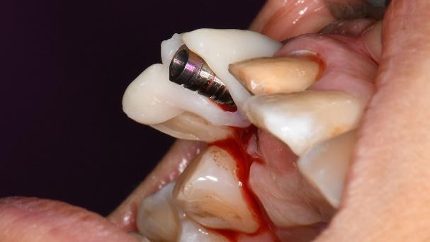
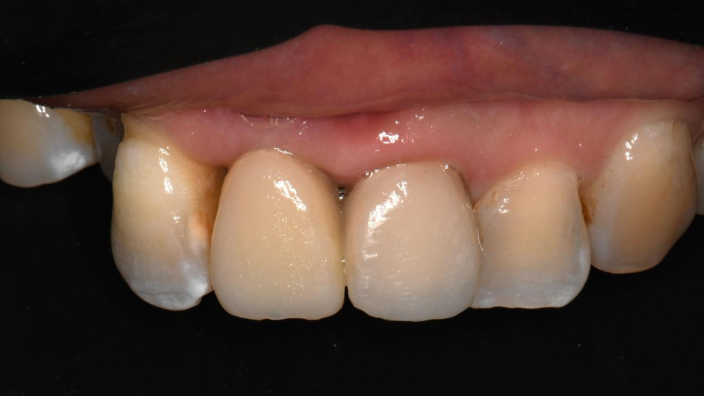
In this case, the anterior teeth were extracted, and the implant body was placed at the target position according to the surgical guide, previously designed. The surgical operation lasted 5 minutes, and the implant position was ideal. The patient was satisfied with the time and experience of the treatment.
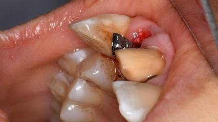
According to the pre-operative scan, the temporary teeth were made in advance, and the immediate post-operative restoration can save a lot of chairside waiting time. The shape of the emergence replicates the neck contour of the natural tooth, creating suitable conditions for permanent restoration in the later stage.
This implantation in anterior teeth was completed by SHINING 3D and Hangzhou Dental Hospital in cooperation, thanks to the support and trust of dentist Dr. Wang Han and technician Zhou Xi.
 ENG
ENG


